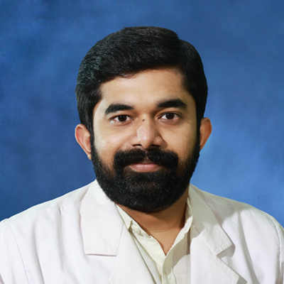
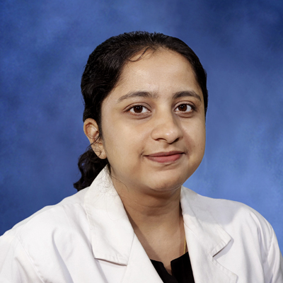
MD - Radiodiagnosis was started in June 2012. Clinical training, lectu...
Read MoreClinical training and lectures are conducted to MBBS students in Radio...
Read MoreInterventional procedures like USG / CT guided FNAC, biopsy, abscess d...
Read More Overview
Department of Radio diagnosis and imaging AMALA IMS, is enriched with most premium models of modern equipment in various modalities. Round the clock services are provided to the patients for routine investigations. Majority of the equipment of various modalities are located in the basement floor of ST.Mary’s block-2 of the Hospital

CT-COMPUTED TOMOGRAPHY:- It combines a series of X-ray images taken from different angles around your body and uses computer processing to create cross sectional images

MRI-MAGNETIC RESONANCE IMAGING:-It uses a powerful magnetic field, radiofrequency pulses and a computer to produce image of the body. MRI does not emit the ionizing radiation.

FLUOROSCOPY:- It is an imaging modality that allows real time X-ray viewing of internal structures by using X-ray tube, image intensifier and TV monitor.

MAMMOGRAPHY :-Mammography unit consists of an X-ray tube and detector to examine the breast

DR-DIGITAL RADIOGRAPHY:- It is an advanced form of X-ray equipment which produces digital radiographic images instantly on a computer.

USG-ULTRASONOGRAPHY :-It is an imaging method that uses high energy sound waves to make echoes that form pictures of internal organs.
MD - Radiodiagnosis was started in June 2012. Clinical training, lecture classes and seminars are conducted regularly as per the NMC norms.
Clinical training and lectures are conducted to MBBS students in Radio Diagnosis department regularly.
Three year diploma in Radiography and Radiotherapy Technology (DRRT), recognized by Govt. of Kerala, conducted by the Directorate of Medical Education (2018 onward).
The Hospital Information System is connected to PACS of Radiodiagnosis Department, to facilitate clinicians and to enhance thesis and research works.
Interventional procedures like USG / CT guided FNAC, biopsy, abscess drainage, pleural fluid / ascites aspiration, catheter placement etc. are also done.
1. All routine and Contrast CT scans.
2. Special investigation procedures like Cerebral, Renal, Pulmonary, Peripheral & Coronary angiograms etc.
3. CT guided interventional procedures like (FNAC/Biopsy/Aspiration, Loopogram, Drainage, Microwave Ablation).
4. Radiotherapy treatment planning.
Radiodiagnosis uses imaging technology to help doctors see what's happening inside the body. The radiology department in Thrissur supports the diagnosis of injuries, infections, tumours, and other health conditions through appropriate imaging investigations.
The major difference between an MRI (Magnetic Resonance Imaging) scan and a CT (Computed Tomography) scan comes down to technology used and what they can detect better than the other. An MRI scan uses magnetic fields and radio waves to create detailed images, particularly of soft tissues. A CT scan uses X-rays and is often useful for examining injuries, bones, organs, and certain internal abnormalities. Your medical doctor will recommend whichever is more suitable.
To examine the abdomen, pelvis, pregnancy-related structures, and certain soft tissues are what Ultrasound scan is commonly used for. It uses sound waves to create images and doesn't involve ionising radiation.
An MRI doesn't usually cause pain. However, the machine can be noisy, and you'll need to stay still during the scan. You should let the professional know beforehand if you are uncomfortable in enclosed spaces. If you are taking an MRI scan in Thrissur, you should visit Amala Hospital for the best possible care.
Preparation depends on the type of scan. Some CT examinations require fasting, follow specific instructions, or contrast material. Inform the radiology team if you have allergies, kidney problems, or a possibility of pregnancy. Visit a reputed CT scan centre in Thrissur like Amala to get the best possible steps before a CT scan.
Yes, X-rays and CT are generally safe as they only involve controlled amounts of radiation. Doctors recommend them when the information they provide is expected to outweigh the associated risks. If you have concerns about radiation exposure, discuss them with your doctor.
Scans can show suspicious growths or changes that need further investigation, but they don't always confirm cancer on their own. In some cases, a biopsy is needed to establish the diagnosis.